Tools, products & open work.
Our methods, in motion.
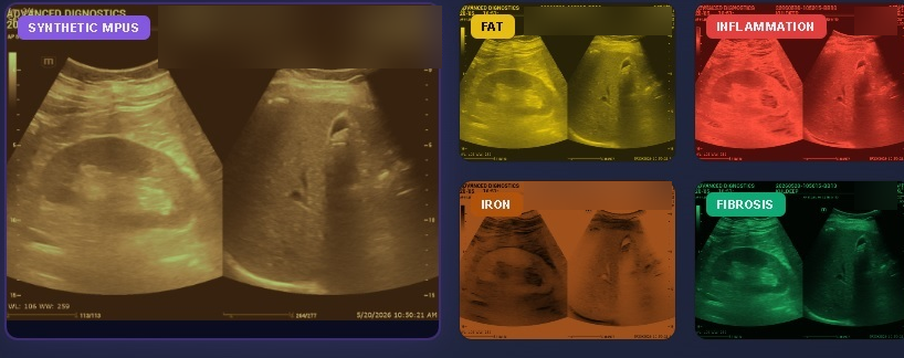
Multiparametric ultrasound for fatty-liver disease
How our AI-assisted multiparametric ultrasound characterises liver fat, inflammation, iron and fibrosis in a single non-invasive read — built to stay accurate even in high-BMI patients where conventional tools struggle.
Is invasive coronary angiography becoming optional?
A walk through coronary CT-FFR: how AI-driven flow simulation adds the functional significance of a stenosis to the anatomy a CT already shows — and models the likely effect of an intervention before it is performed.
The case for CT-first coronary assessment
A silent, motion-graphic primer on where coronary CT-FFR fits in stable chest-pain pathways — the quick visual companion to the narrated walk-through.
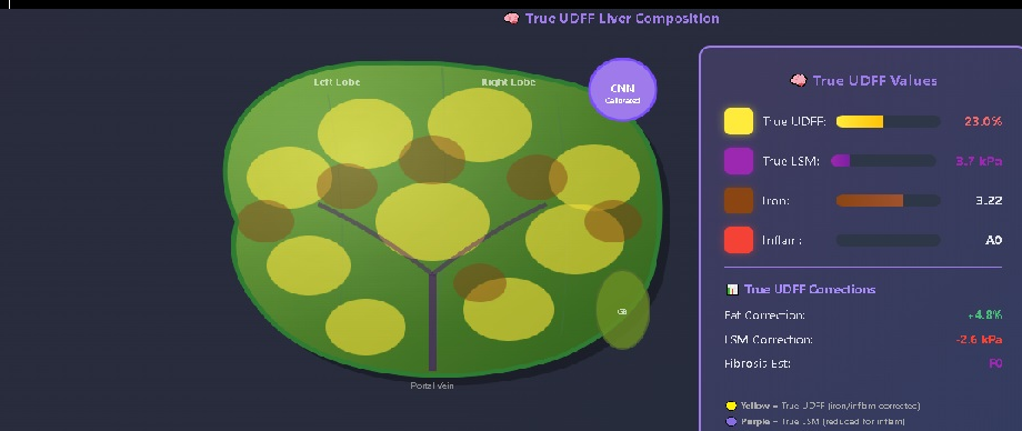
FibroPlus — Multiparametric Liver Scan
MAFLD is now a global epidemic, affecting more than 30% of the population. It is both a cardiometabolic risk factor and a precursor to MASH and progressive fibrosis. Conventional tools are often inaccurate in high-BMI patients — producing false positives and negatives at high cost — and miss the contributions of inflammation and iron. FibroPlus is a unique AI-assisted multiparametric ultrasound that overcomes these limitations, supporting the clinical management and classification of MAFLD patients.
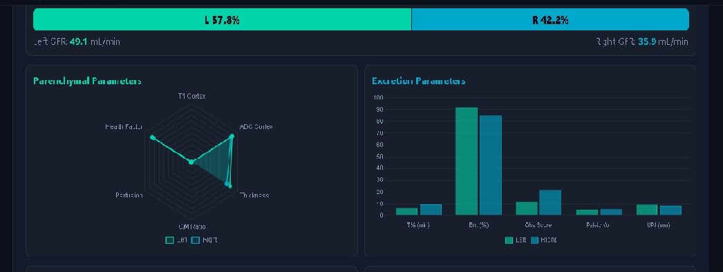
Non-Contrast MRI — Renal Parenchyma & Excretion Analysis
A novel method for assessing renal parenchymal health that simulates excretory analysis — comparable to a DTPA urogram — without isotopes or contrast agents, giving a reliable prediction of split renal function and overall kidney health.
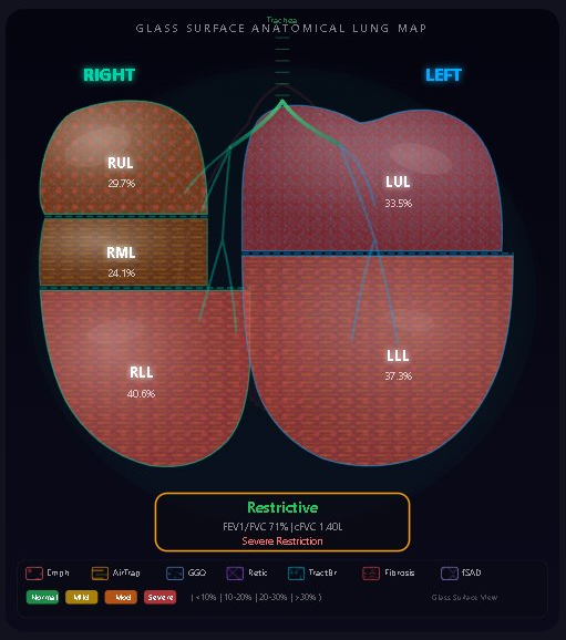
PULMO — Quantitative Lung CT with Spirometric Assessment
An AI-assisted tool that pairs CT lung function with anatomical evaluation across full inspiration and expiration. It identifies and classifies the type of respiratory disease while quantifying the degree of ILD and emphysema.
CT-FFR with AI Flow Simulation
Coronary CT is a well-accepted tool for evaluating stable CAD. Adding FFR captures the functional significance of a stenosis, and real-time AI flow simulation can model the effect of an intervention before the procedure is chosen.


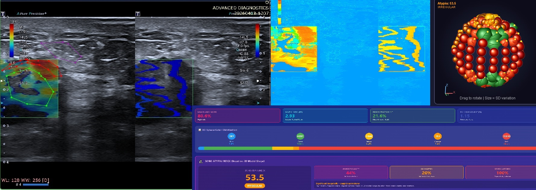
SonoHistology of Nodules — Breast, Prostate & Thyroid
Ultrasound-based tissue characterisation of nodules in the breast, prostate and thyroid. It combines elastography with quantitative imaging features to help stratify lesions non-invasively and inform the decision to biopsy.
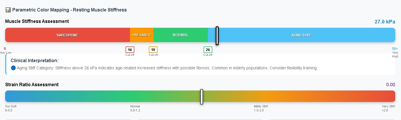
Sarcopenia — Non-Invasive Evaluation
Muscle loss is both physiological and pathological, and is closely linked to morbidity and longevity. Using a dedicated algorithm, this tool distinguishes age-related muscle loss from true sarcopenia and quantifies its severity.
Q-FFR — for Coronary Catheter Angiography
Derives FFR directly from coronary catheter angiography images to assess the functional relevance of a stenosis, and helps classify the pattern of ischaemic lesions as focal or diffuse.

| Territory | Worst FFR | Verdict |
|---|---|---|
| LAD | 0.87 | Defer |
| LCx | 0.82 | Defer |
| RCA | 0.97 | Defer |